During a Specialty Contact Lens Consultation at In Focus, Dr. Morrison often explains the difference between scan-designed scleral lenses and conventional scleral lenses to patients when they are deciding on lens options. Many patients are candidates for both types of sclerals, and in most cases, one type will be more beneficial than the other, like the patient below.
This patient has keratoconus with 60.00 D K values (steepness of cornea) which is much steeper than average (average D K values fall between 50.00 – 55.00).
Their vision with and without glasses was counting fingers in front of face, which is considered to be quite low.
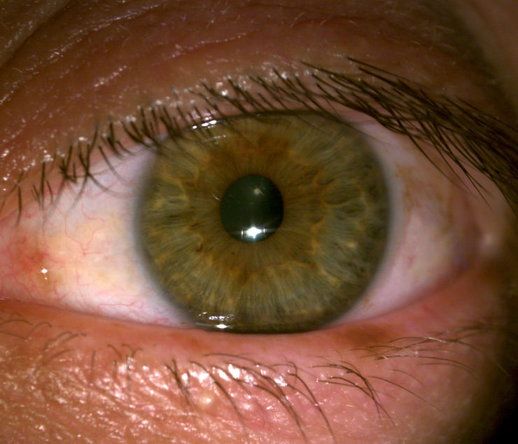
In the picture below, we can also see that this patient has bumps located nasally and temporally on their eye called pinguecula, which are small, raised, white- or yellow-colored growths made of protein, fat, or calcium deposits. These are very common and are usually due to exposure to the UV rays of the sun.
While this patient could use either type of scleral lens with success, Dr. Morrison chose scan-designed lenses for this case in order to create more grooves over the two areas with pinguecula to more easily match the cornea steepness.
With more advanced designs of scleral lenses (scan and mold/impression-based designs), the grooves to accommodate the pinguecula are already created in the lens since they are detected by eye scans or eye molds. With conventional sclerals, we have to make on-the-eye measurements to create little channels for these mounds of tissue, which isn’t as precise and means the process takes a little longer to accurately match the surface of the patient’s eye.
Great news for this patient: when they tried the lenses, their vision increased to 20/30! We are excited for them to try on their first pair in a few weeks.

#ScleralLenses #Keratoconus #ScanDesignedSclerals #MoldDesignedSclerals #EyeScan #EyeMold #pinguecula #ConventionalSclerals #Sclerals #CustomDesign #CustomLens #SpecialtyContactLenses #LensDesign #ContactLensSpecialist #Optometry #LensSpecialist

